Testing a mattress for free before trying it out is a fantastic opportunity. Here’s how you can try out online mattresses in person before buying.
Blog > Electroencephalography (EEG) for Sleep Disorders Diagnosis 2024
Blog > Electroencephalography (EEG) for Sleep Disorders Diagnosis 2024
Electroencephalography (EEG) is a non-invasive and non-harmful diagnostic process used for recording abnormal electrical activity in the brain. The results taken through this technique are called Electroencephalograms.
All communications within our body occur in the form of electrical impulses. In an EEG, the signals are detected in the form of waves. Any abnormal activity is readily detectable because of the changes in waves.
Around the world, EEG is being used as a diagnostic tool for brain-related diseases such as epilepsy, tumors, head injuries, and Alzheimer’s. Even experts who sedate you before surgery want to ensure the correct dose of anesthesia during a critical surgery. Many doctors also recommend an EEG to patients who have sleep-related issues.
All the electrical activities are detected through a set of electrodes placed on your scalp surface. Electrodes are the conductors through which electric waves travel responsible for transferring brain waves to EEG machines.
The process of electroencephalography takes about 20 to 30 minutes, in some cases, test may take about an hour or two. Once the recording begins you will be asked to stay still while the technician will monitor you through a window.
During the test, your technician may ask you to breathe deeply or rapidly to produce prominent brain wave activity. Normally you will be asked to just close your eyes but if you are being tested for any sleep disorder, the test can be done while you remain asleep.
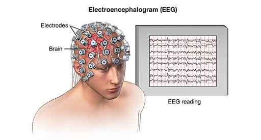
As discussed earlier, the most common tests conducted by neurologists to detect brain wave activity is called EEG. Let’s discuss few types of EEG tests.
Multiple types of EEG test are done for different evaluations, such as, standard EEG, ambulatory, video, invasive, neurofeedback, polysomnography. However, most commonly used are sleep EEG, sleep deprived EEG and polysomnography.
One of the most common EEG tests is sleep-deprived EEG conducted by neurologists to recognize any abnormal brain wave activity and sleep disorders. Sleep-deprived EEG tests are most likely taken when a patient is asked to stay awake a night before the test and avoid any drinks with caffeine.
Sleep EEG tests are performed to detect any abnormal brain wave activity while a person is asleep. In most cases, the quality of your sleep cycle is identified via a Sleep EEG test.
Polysomnography, a type of sleep study is an EEG test to identify any sleep disorders. Polysomnography is performed either at the hospital or a sleep clinic; asleep technician studies your sleep cycle stages, brain wave activity, eye movements in detail to detect any sleep disorders.
Detection of human brain activity via electroencephalography (EEG) is being used for 80 years. Learn about the history of Electroencephalography (EEG).
EEG remains one of the oldest and most reliable methods to record brain wave activity and detect any sleep disorders for further treatment.
The history of electroencephalography began from 1929 when a German psychiatrist Hans Berger discovered this neurological tool. Hans Berg recorded some instincts of brain waves from a patient’s scalp; and it was the first time he found about nerve recordings.
Berger’s discovery was later confirmed in 1934 at Harvard in the United States and England. Further progress was made in EEG development. A study reported by Harvard in 1935 stated, EEG is a useful neurological tool for the detection of normal/abnormal brain wave activity, seizures, strokes and disorders.
Recently in October 2018, an experiment was conducted by neurologists, where they connected the brains of three people to share and exchange their thoughts.
The experiment was done via electroencephalograms (EEGs) to detect individual brain wave activity and transcranial magnetic stimulation (TMS). Researchers revealed, findings from EEG can be used to connect multiple minds and share their thoughts among each other.
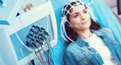
Preparing for an EEG test recording
An EEG is a non-harmful procedure, thus no risks are associated with it. Learn more about EEG and whether it involves risks for better understanding.
EEG test does not involve any risks. It is completely harmless and painless. There are zero radiations or medications involved; the analysis is merely performed through the topical application of electrodes on the scalp.
First, the sleep technician will ask you to lay down as he marks where the electrodes will be placed on your scalp. A gel is then rubbed on those spots for better reading, and further, the electrodes are placed on your scalp.
During the test, the technician may ask you to close your eyes and stay still. Besides the basic application, no other procedures are done.
EEG tests are entirely safe and does not involve any risks. However certain factors might affect EEG test readings which includes anxiety issues, medical conditions and so on.
Factors that influence EEG readings include,
Your doctor and technician will usually tell you what to do before an EEG test and how to prepare for it. To get an idea, here are few of the steps you must take before the test.
EEG is profoundly used to identify and diagnose any sleep disorders. Explore how EEG is helpful in the diagnosis of sleep disorders.
Your doctor might suggest a detailed EEG sleep study or polysomnography, in order to get an idea about your brain activity inside. A polysomnography includes a head EEG among other tests, such as study of body movements or blood oxygen levels and heart rate monitoring.
As discussed earlier, the EEG process starts with the placement of electrodes on the scalp using a mild glue. Electrodes are tiny metal discs measuring the electrical signals or voltages in the brain. Each of the discs is wired up to a machinery to display the results. The report can either be printed in the form of a graph or viewed on the computer screen.
The technique monitors and displays the anomalies in your brain impulses which may be causing a sleep issue. Unusual body movements or voltage spikes are observed during the EEG of sleep stages, including NREM and REM, to detect the abnormal behavioral patterns you present while slumbering.
The final EEG results are interpreted by a clinician or a specially trained sleep specialist. They analyze the type or cause of the disturbance leading to uninterrupted or restless sleep.
You may be tested at a sleep lab, a specialized environment with multiple machines to assess how you rest or the electrical activity that happens in your brain during sleep. In other cases, you may be given portable home EEG sleep monitor to conduct the sleep tests at the comfort of your house.
Let’s explore how EEG can be used to detect the 6 most popular sleep disorders.
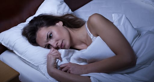
Sleep disorders can be identified via EEG readings
Sleep deprivation is a condition due to inadequate sleep, usually caused by sleeplessness or disorder of circadian rhythms. Commonly, there are two kinds of sleep deprivation, chronic and acute.
Chronic sleep deprivation is long periods of sleep deprivation, profoundly affecting your mental and physical health.
It also decreases your productivity and performance. Some common symptoms are weight loss, weight gain or fatigues.
Acute sleep deprivation is a shorter period with disrupted or the lack of sleep. Some common symptoms for the following kind are, mood disorders, yawning, lack of concentration, dizziness.
However, as the deprivation period is short, symptoms do not stay for long either.
EEG is found to be extremely helpful for sleep-deprived individuals. The symptoms you experience generate brain waves which can be seen through EEG. Most of the times, people fall asleep during the EEG test, the unseen brain waves can also be calculated through it.
Any risks of epilepsy or seizure can be detected with the help of sleep deprivation EEG and early treatments can be taken.
Before the test, you are required to stay awake for 24 hours, also not to consume anything containing caffeine.
Obstructive sleep apnea (OSA) is a chronic condition in which the affected person experiences airflow blockage while sleeping due to sudden relaxation of the throat muscles causing passage hindrance. Two basic types of sleep apnea exists, including obstructive sleep apnea and central apnea, however, 80% of individuals suffer from obstructive sleep apnea.
Sleep apnea occurs when you actually stop breathing for about 10 seconds, but suddenly your brain waves alert you to wake up and take a deep breath. Some individuals experience sleep apnea about 30 times per night.
Regardless whether you are sleeping for 6-8 hours, if you have a chronic sleep disorder of sleep apnea, you might still feel tired in the morning as you are waking up plenty of times at night in the middle.
Research shows, an EEG examination among epilepsy patients can be helpful in detecting early symptoms of sleep apnea. An EEG Sleep Apnea test on OSA patients can capture alterations in brain waves while sleeping due to interrupted respiration.
Parasomnia is a sleep disorder characterized by abnormal behavioral activities during sleep, such as night terrors, sleepwalking, sleep talking, etc. This disorder may or may not occur along with epileptic seizures.
According to a 2014 study by MK. Pavlova, S. Yazdani, and EJ. Bubrick of Harvard Medical School Department of Neurology, multiple patients, with no history or risk of epilepsy, suddenly exhibited epileptic brain wave patterns in an EEG.
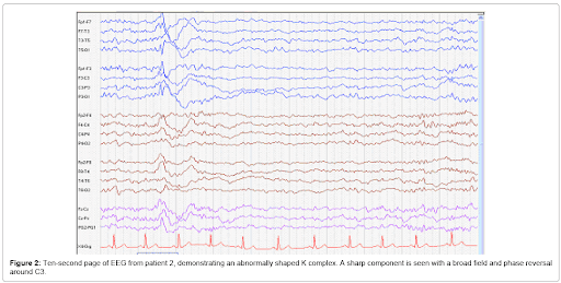
Unusual Epileptic Spikes Indicating Parasomnia
The interesting part about this is that the parasomnia EEG spikes during sleep are usually different from those observed in epileptic cases.
The scientists concluded that the sleep waves associated with cortical irritability (wave patterns indicating seizures), triggering in patients with no history of epilepsy, may become a useful way to diagnose parasomnia through EEG.
Insomnia is one of the most highly diagnosed sleep disorders globally. It refers to the loss of sleep or inability to fall asleep at night.
Insomnia is all about trouble in falling asleep and sleep disruptions. Regardless of how much you workout or how appropriately you eat, if you have a poor sleep quality, you will never feel relaxed or active once you wake up.
In today’s world, insomnia and sleep deprivation are often confused with one another. However, both of them are completely distinctive. Insomnia is about inability to fall asleep, regardless of having opportunity ; whereas, sleep deprivation is a state of poor sleep quality, with lack of opportunity.
Per the American Academy of Sleep Medicine (AASM), over 30% of the American adults show mild insomnia symptoms whereas around 10% have been diagnosed with chronic insomnia.
A sleep EEG is pretty helpful in diagnosing insomnia. During this condition, the human brain emits slow waves, and the central nervous system (nerves in the brain and spinal cord) experiences enhanced hyperarousal, leading to difficulty in falling asleep.
Hence, the slow wave activity on EEG is a clear indication of insomnia, which goes to show that this technique is critical in assisting with sleep disorders diagnosis.
Sleep paralysis is perhaps the scariest of all sleep disorders. It is a condition in which you are consciously laying on the bed, yet you experience heaviness on the chest, difficulty in breathing, or are unable to speak and move.
Sleep paralysis is somehow associated with psychiatric problems, usually occurs when your sleep cycle is not properly conducted or you are in between the stages of sleep and wakefulness. Study shows, sleep paralysis might occur one or two times per night, depending on psychiatric or physical attributes of a person.
Originally, two basic kinds of sleep paralysis exist, known as hypnagogic/ predormital sleep paralysis and hypnopompic/postorbital sleep paralysis.
To get a clear idea about sleep paralysis, you might want to watch this video. SPOILER ALERT – it’s scary!
Scientists use advanced EEG techniques, coupled with fMRI, to detect the occurrence of sleep paralysis. Most people with sleep paralysis, show reactions in certain regions of the brain that are otherwise shown resting in the REM sleep stage EEG. Thus, an EEG report is helpful in identifying sleep paralysis onset.
Lastly, if the sleep paralysis symptoms are initial and not quite severe, you can opt for basic treatments such as opening and moving your eyes around for comfort, sleeping in a regular pattern, exercising regularly or eating healthy etc.
People who suffer from narcolepsy have sudden involuntary episodes of sleepiness, both during quiet time or while performing some activity, such as working in an office or driving a car.

Narcolepsy brain waves are similar to that of epilepsy. Some individuals also have cataplexy attacks (sudden loss of muscle control triggered due to a strong emotion) along with narcolepsy, which further clarifies the specific type of sleep disorder.
Usually, narcolepsy EEG results show increased blinking of eyes or body movements, while the patient is undergoing polysomnography. The sleep technicians can instantly find the abnormal sleep EEG spikes in the report caused due to these movements in the early stages of the sleep cycle, confirming the symptoms of narcolepsy.
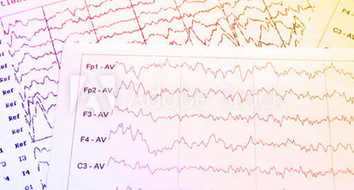
Here are some top interesting facts you probably didn’t know about EEG.
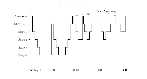
Sleep patterns travel in the form of brain activity
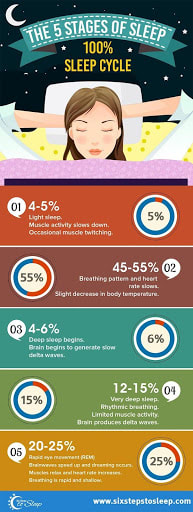
Your body and mind travels through five basic stages during sleep. Sleep cycle stages are typically categorized into two types, NREM (non- rapid eye movement) and REM (rapid eye movement).
Sleep cycle and patterns travel in the form of brain wave activity, which can be identified easily with electroencephalography (EEG) readings.
Sleep researchers usually monitor brain wave activity and changes in it via EEG recordings. Moreover, any kind of sleep disorder is easily detectable during sleep cycle EEG.
Do you know your brain contains millions of nerves, known as neurons? Neurons communicate with each other that creates electrical activity into your brain.
These electrical activity are called brainwaves, which can be detected via EEG – electroencephalography machine. Following brain waves can be detected with the help of EEG machine. Let’s discuss about the brain waves for proper sleep cycle, so if you notice any changes or abnormalities, there might be a presence of any sleep disorder!
Delta waves for any healthy sleep cycle are highest in amplitude with slowest waves. They usually appear during Stage 3 and Stage 4 and appears to be the prominent brain wave in infants. Usually, delta waves own a frequency of 3 Hz or lower.

Theta waves are pretty dominant in children with 13 years or more but quite unusual in adults. These waves own a frequency of 3.5 to 7.5 Hz for normal sleep cycle detection. Typically, there is presence of theta waves in disorders known as metabolic encephalopathy or hydrocephalus.

Alpha waves are one of the most dominant waves in adults, usually detected in high amplitude in your posterior areas with a frequency of 7.5 and 13 Hz. Appearance of Alpha waves occurs when you close your eyes and goes away when you wake up.

Alpha waves in normal EEG reading
Beta waves appear with the highest and fastest amplitude of 14 and greater Hz. They appear to be the highest when a person is wide awake, commonly on both sides of the symmetrical distribution.

In conclusion, sleep EEG testing is an excellent, highly accurate way of diagnosing a variety of sleep disorders. The result interpretations by trained sleep technicians eventually assist doctors in preparing an effective medication or care plan for the patients, decreasing health risks due to incorrect sleep diagnosis.

 Showrooms
About The Author
Dustin Morgan
Sleep Expert & Store Manager
Dustin Morgan is the Chief Mattress Analyst and Sleep Technology Expert at SleePare. He combines his computer science background with his passion for sleep innovation. With personal experience testing over 200 mattresses, Dustin offers unmatched insights into finding the perfect sleep solution for various needs. His work focuses on delivering honest and detailed comparisons and advice to help individuals achieve their best sleep. When he is not exploring the latest sleep technology, Dustin enjoys spending time in the great outdoors and gaming in his Virginia home.